Diagram Of Shoulder : Free Shoulders Cliparts Diagram Download Free Shoulders Cliparts Diagram Png Images Free Cliparts On Clipart Library - The acromioclavicular (ac) joint connects the upper part of the shoulder blade to the collarbone, or clavicle.
Diagram Of Shoulder : Free Shoulders Cliparts Diagram Download Free Shoulders Cliparts Diagram Png Images Free Cliparts On Clipart Library - The acromioclavicular (ac) joint connects the upper part of the shoulder blade to the collarbone, or clavicle.. There are two joints within the shoulder that can be affected by osteoarthritis. This is where the humerus (arm bone) meets the scapula (shoulder blade). Common rotator cuff injuries include rotator cuff tendonitis and rotator cuff strain, which is a partial or complete tear of the rotator cuff. A diagram of an anatomic shoulder replacement—the plastic socket replaces the cup of the scapula (shoulder blade). Pronate your wrist so the palm of your hand faces down to the floor (as if you were trying to empty a glass of water).
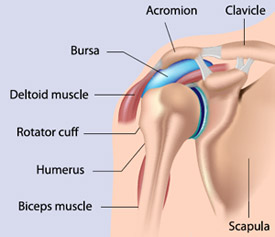
The shoulder is a complex combination of bones and joints where many muscles act to provide the widest range of motion of any part of the body. There are 10 muscles and 11 shoulder tendons related to shoulder mobility. The shoulder girdle includes three bones—the scapula, clavicle and humerus. Numerous muscles help stabilize the three joints of. A second joint in the shoulder is the junction of the collar bone with the shoulder blade, called the.
Understanding Your Shoulder Injury Sunnybrook Hospital from sunnybrook.ca The glenohumeral joint is a joint where the greater tubercle (humeral head at the top of the arm bone) meets the shoulder socket of the scapula, called the glenoid cavity or glenoid fossa. Inside the shoulder there are three joints; A second joint in the shoulder is the junction of the collar bone with the shoulder blade, called the. The shoulder has about eight muscles that attach to the scapula, humerus, and clavicle. The humeral head is the ball side. Inability to carry objects or use your arm. Make sure the arm is positioned 30 degrees forward as you raise it (see diagram) 5. They may use shoulder joint diagrams to understand shoulder joint anatomy.
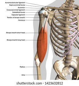
Bones in shoulder, ligaments of the shoulder joint, parts of the shoulder joint, shoulder anatomy, shoulder joints and muscles, shoulder structure anatomy, shoulder tendon anatomy, shoulder tendons ligaments, human muscles, bones in shoulder, ligaments of the shoulder joint, parts of.
It is one of the most mobile joints in the human body, at the cost of joint stability. The acromioclavicular (ac) joint connects the upper part of the shoulder blade to the collarbone, or clavicle. In this episode of eorthopodtv, orthopaedic surgeon randale c. Shoulder skeleton diagram with head and deltoid tubercle of humerus, scapula skeletal structure anatomy of neck and shoulder stock illustrations. A guide to understand shoulder joint with diagram. What are common rotator cuff injuries? The primary function of the shoulder girdle is to give strength and range of motion to the arm. The bones of the pectoral girdle (clavicle and scapula) provide increased mobility to the. These muscles form the outer shape of the shoulder and underarm. Numerous muscles help stabilize the three joints of. Some signs that you should be seen by a doctor include: The shoulder joint (glenohumeral joint) is a ball and socket joint between the scapula and the humerus.it is the major joint connecting the upper limb to the trunk. An injury that causes joint deformity.
Shoulder pain that occurs at night or while resting. The muscles in the shoulder aid in a wide. Shoulder skeleton diagram with head and deltoid tubercle of humerus, scapula skeletal structure anatomy of neck and shoulder stock illustrations. Ebraheim's educational animated video describes muscle anatomy of the shoulder girdle and anatomy of the shoulder joint.anatomy of the shoulder muscles a. It is an extremely mobile joint, in which stability has been sacrificed for mobility.
Shoulder Anatomy Labeled Images Stock Photos Vectors Shutterstock from image.shutterstock.com The shoulder joint is an active joint that assists the forward and backward movement of the shoulder. There are 10 muscles and 11 shoulder tendons related to shoulder mobility. The most flexible joint in the entire human body, our shoulder joint is formed by the union of the humerus, the scapula (or shoulder blade), and the clavicle (or collarbone). Ebraheim's educational animated video describes muscle anatomy of the shoulder girdle and anatomy of the shoulder joint.anatomy of the shoulder muscles a. It is one of the most mobile joints in the human body, at the cost of joint stability. The glenohumeral joint, the acromioclavicular joint (a/c joint) and the sternoclavicular joint. This is the smallest rotator cuff muscle. It is an extremely mobile joint, in which stability has been sacrificed for mobility.
The acromioclavicular joint is where the acromion, part of the shoulder blade (scapula) and the collar bone (clavicle) meet.
Two joints are at the shoulder. Its main job is to assist with rotation of the arm away from the body. The supraspinatus, the infraspinatus, the teres minor and the subscapularis. The shoulder muscles and shoulder tendons involved with shoulder mobility include the four rotator cuff muscle and tendon pairs: The large bone in the upper arm is called the humerus. The top of the humerus is shaped like a ball. The shoulder joint is formed where the humerus (upper arm bone) fits into the scapula (shoulder blade), like a ball and. Ebraheim's educational animated video describes muscle anatomy of the shoulder girdle and anatomy of the shoulder joint.anatomy of the shoulder muscles a. 17 photos of the diagram of shoulder muscles and tendons. The shoulder is not a single joint, but a complex arrangement of bones, ligaments, muscles, and tendons that is better called the shoulder girdle. The glenohumeral joint is a joint where the greater tubercle (humeral head at the top of the arm bone) meets the shoulder socket of the scapula, called the glenoid cavity or glenoid fossa. Raise arm upward to just below shoulder height as shown 4. The muscles in the shoulder aid in a wide.
Some signs that you should be seen by a doctor include: The muscles in the shoulder aid in a wide. The shoulder girdle includes three bones—the scapula, clavicle and humerus. The glenohumeral joint connects the shoulder socket, or glenoid, which extends from the shoulder blade, to the arm bone, or humerus. The shoulder isn't just one bone, it's actually made up of three different bones and various tendons, ligaments, and muscles.the three bones located in the shoulder are the humerus, the scapula, and the clavicle.
Muscle Diagram Shoulder Koibana Info Arm Muscle Anatomy Human Body Anatomy Shoulder Anatomy from i.pinimg.com There are two joints within the shoulder that can be affected by osteoarthritis. Shoulder structure anatomy 17 photos of the shoulder structure anatomy bones of the shoulder joint, parts of the shoulder joint, shoulder bone structure, shoulder bones, shoulder diagram, shoulder parts of the body, shoulder tendon anatomy, shoulder tendons ligaments, hand, bones of the shoulder joint, parts of the shoulder joint, shoulder. A guide to understand shoulder joint with diagram. Is the wear and tear of shoulder cartilage until bare bone is exposed. The glenohumeral joint is a joint where the greater tubercle (humeral head at the top of the arm bone) meets the shoulder socket of the scapula, called the glenoid cavity or glenoid fossa. Some signs that you should be seen by a doctor include: Make sure the arm is positioned 30 degrees forward as you raise it (see diagram) 5. An injury that causes joint deformity.
Common rotator cuff injuries include rotator cuff tendonitis and rotator cuff strain, which is a partial or complete tear of the rotator cuff.
The glenohumeral joint connects the shoulder socket, or glenoid, which extends from the shoulder blade, to the arm bone, or humerus. The humeral head is the ball side. This is the smallest rotator cuff muscle. On the left is a standard (anatomic) shoulder arthroplasty. These are located in the shoulder blade area, and each related tendon also attaches to the humerus. Numerous muscles help stabilize the three joints of. Shoulder pain that persists beyond a few days. The supraspinatus is located on the upper part of the shoulder joint and is involved in abduction (arm raising). The shoulder has about eight muscles that attach to the scapula, humerus, and clavicle. Its main job is to assist with rotation of the arm away from the body. Common rotator cuff injuries include rotator cuff tendonitis and rotator cuff strain, which is a partial or complete tear of the rotator cuff. This is the main muscle that lets you rotate and extend your shoulder. Shoulder skeleton diagram with head and deltoid tubercle of humerus, scapula skeletal structure anatomy of neck and shoulder stock illustrations.


0 Komentar